Diabetic Retinopathy:
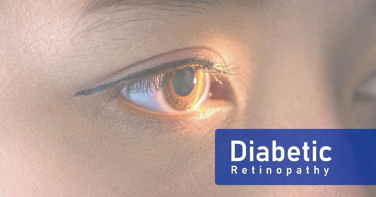
Diabetic Retinopathy, also referred to as Diabetic Eye Disease (DED), is a medical condition in which damage occurs to the retina due to diabetes mellitus. This is one of the leading causesof blindness in developed countries. It is caused by the damage to the blood vessels in the retina, the light-sensitive tissue at the back of the eye. Figure 1 shows the images of healthy vision and diabetic retinopathy patent's vision.
The rating is based on the following symptoms. A few microaneurysms without any other abnormalities indicate mild DR. Cotton-wool spots and hemorrhages indicate moderate DR.
The presence of neovascularization of the disc or elsewhere or vitreous hemorrhage indicates proliferative DR. This is a method that detects important pixels by making a backward transition in a neural network. In the reverse shift, the neurons that contribute most to the upper layer get the most communication.
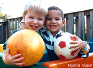
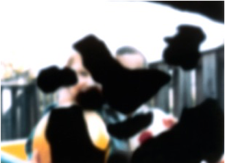
Fig. 1. Normal vision (left) and what DR patient sees (right) (source: National Eye Institute, NIH)
Why does it occur?
Diabetic retinopathy affects up to 80% of people who have had diabetes for 20 years or more. At least 90% of new cases could be reduced with proper treatment and monitoring of the eyes. The longer a person has diabetes, the higher their chances of developing diabetic retinopathy.
Treatment for diabetic retinopathy:
Treatment is based on the cause of the retinopathy and may include laser therapy to the retina. Laser photocoagulation is the standard treatment for many types of retinopathy. Evidence suggests that laser treatment is generally safe and improves the visual symptoms of sickle cell disease and diabetic retinopathy. In recent years targeting the pathway controlling vessel growth or angiogenesis has been promising. Vascular endothelial growth factor (VEGF) plays a vital role in promoting the formation of new blood vessels. Using anti-VEGF drugs (antibodies to sequester the growth factor), research has shown a significant reduction in the extent of vessel outgrowth. Low-quality evidence supports anti-VEGF antibodies, such as bevacizumab or pegaptanib, which seem to improve outcomes when used in conjunction with laser therapy to treat retinopathy of prematurity; longer-term systemic effects are not known, however. The evidence is poorer for the treatment of diabetic retinopathy. The use of anti-VEGF drugs did not appear to improve outcomes compared to standard laser therapy for diabetic retinopathy.
diabetic retinopathy vs. normal eye
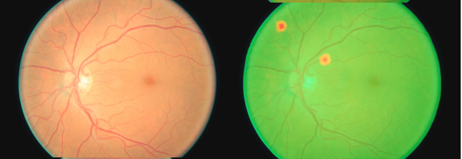
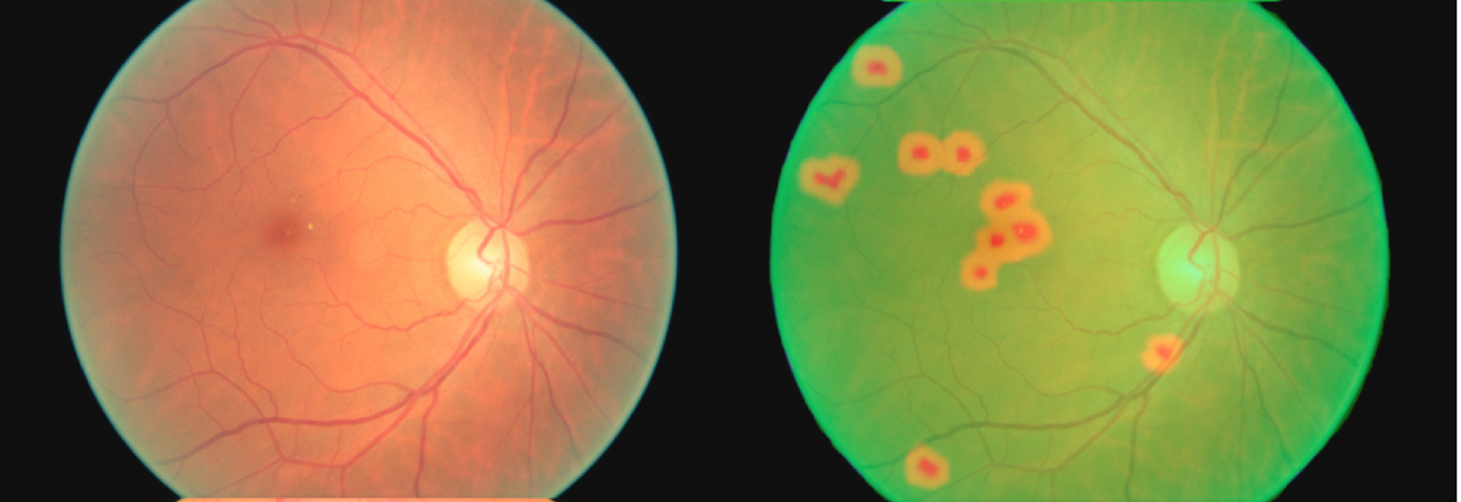
Five stages of DR development using the Kaggle dataset example. (A) Normal eye, (B) Mild DR, (C) Moderate DR Eye, (D) Severe DR (proliferative), and (E) Proliferative DR. Severe and Proliferative DR include the heat map of the affected areas (hemorrhages, exudates, microaneurysms, neovascularization, etc.).
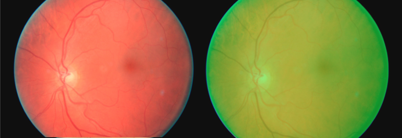
No DR (normal eye) Mild DR Moderate DR eye
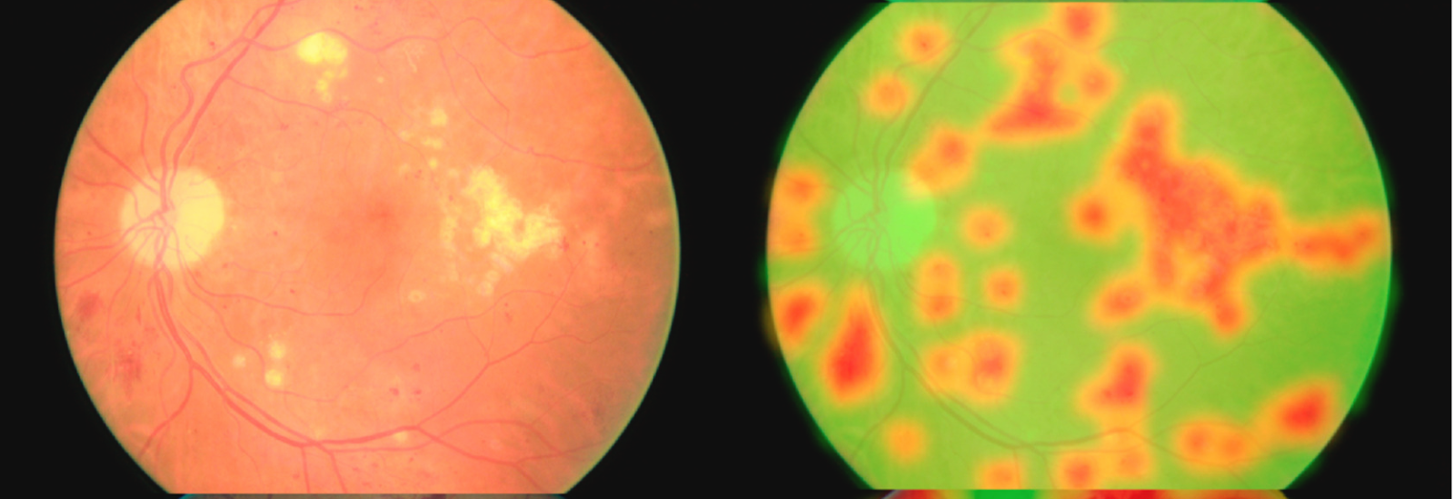
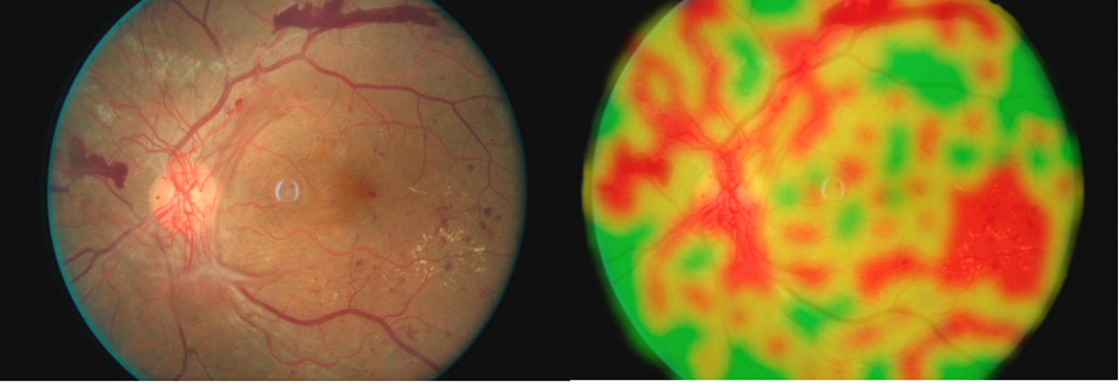
Severe DR (proliferative) Proliferative (severe non-proliferative eye)
Diabetic Retinopathy Screening
typically have no symptoms – it's often too late to reverse them by the time patients visit Ophthalmologist. Early detection & preventive care are essential: there's no treatment after progression. Conventional (manual) screening depends on PCP referral to specialists; many patients don't comply, and everyone faces delays due to ophthalmologist access issues. The majority of the at-risk population is not being served until too late.
AI based Diabetic Retinopathy (DR) Screening
What is AI based DR screening: AI based Diabetic Retinopathy screening system is automatic Using a computer algorithm to analyze the images and detect the severity of DR.
What is the benefit of AI based DR Screening ?
Early detection is key to prevention, which could be achieved effectively with a fully automated screening tool performing well on clinically relevant measures in primary care settings. We have built a tool based on artificial intelligence on a cloud platform for extensive DR testing as referenceable or not recommended. This paper aims to validate this tool built using deep learning-based techniques.
The cloud-based software for testing the automated system compared to expert ophthalmologists' evaluations of referable DR. Measures used as the area under the curve (AUC), sensitivity, and specificity of the screening model concerning professional graders.
Over 98% of cases, and nearly 100% of the normal cases are correctly identified by our system (more than 100K images).
|
o Metric o |
o
o DR o |
o
o AMD o |
o
o Glaucoma o |
o
|
o Sensitivity o |
o
o 0.976 o |
o
o 0.989 o |
o
o 0.83 o |
o
|
o Specificity o |
o
o 0.994 o |
o
o 0.995 o |
o
o 0.94 o |
o
|
o AUC o |
o
o 0.99 o |
o
o 0.99 o |
o
o 0.93 o |
o
The screening system achieved a high sensitivity of 99.21% and a specificity of 97.59% on the Kaggle test dataset with an AUC of 0.9992. The system was also externally validated in Messidor-2, where it achieved a sensitivity of 97.63% and a specificity of 99.49% (AUC, 0.9985). On primary care data, the sensitivity was 92.3% overall (12/13 referable images are correctly identified), and overall specificity was 94.8% (233/251 non-referable images).
How iPredict does this automatic screening: is an artificial intelligence-based tool on a cloud-based platform for large-scale screening of DR as referable or non-referable. This paper aims to validate this tool built using deep learning-based techniques.
Accuracy: xx
Early detection can save millions of people's eyesight and significantly reduce medical costs. iHealthScreen iPredict is a hipaa compliant Ai powered screening system serving diabetic retinopathy eye exams.
Comments
1
1
1
1
1
1
1
1
1
1
1
1
1
1
1
1
1
1
1
1
1
1
1
1
1
1
1
1
1
1
1
1
1
1XnziMnp4
1
-1 OR 5*5=25 --
-1 OR 5*5=25
-1' OR 5*5=25 --
-1" OR 5*5=25 --
-1' OR 5*5=25 or 'JknCj00n'='
-1" OR 5*5=25 or "2l8svByJ"="
1*if(now()=sysdate(),sleep(15),0)
10'XOR(1*if(now()=sysdate(),sleep(15),0))XOR'Z
10"XOR(1*if(now()=sysdate(),sleep(15),0))XOR"Z
(select(0)from(select(sleep(15)))v)/*'+(select(0)from(select(sleep(15)))v)+'"+(select(0)from(select(sleep(15)))v)+"*/
1-1; waitfor delay '0:0:15' --
1-1); waitfor delay '0:0:15' --
1-1 waitfor delay '0:0:15' --
1HQ8nlGVK'; waitfor delay '0:0:15' --
1-1 OR 520=(SELECT 520 FROM PG_SLEEP(15))--
1-1) OR 26=(SELECT 26 FROM PG_SLEEP(15))--
1-1)) OR 578=(SELECT 578 FROM PG_SLEEP(15))--
1ijknx1Qe' OR 869=(SELECT 869 FROM PG_SLEEP(15))--
1bGKo0YkE') OR 198=(SELECT 198 FROM PG_SLEEP(15))--
1oMTjS9h9')) OR 886=(SELECT 886 FROM PG_SLEEP(15))--
1*DBMS_PIPE.RECEIVE_MESSAGE(CHR(99)||CHR(99)||CHR(99),15)
1'||DBMS_PIPE.RECEIVE_MESSAGE(CHR(98)||CHR(98)||CHR(98),15)||'
1
1'"
@@ctRJX
(select 198766*667891)
(select 198766*667891 from DUAL)
1
1
1
1
1
1
1
1
1
1
1
1
1
1
1
1
1
1
1
1
1
1
1
1
1
1
1
1
1
1
1
1
1
1
1


Very informative.